This male patient had a history of trauma followed by scrotal pain and swelling. Ultrasound shows bilateral tunica vaginalis hydrocele with multiple septae with the left side being more affected.
The 3-D ultrasound images below show fine detail of the septations within the scrotal hydrocele.
with the history of from, the possibility of this fluid collection being a hematocele should be considered.

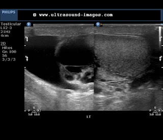


The left testes also shows tubular ectasia of the rete testes- in the 3-D ultrasound image below:


Visit: http://www.ultrasound-images.com/scrotum/#Hydrocele-%20B-mode%20grey%20scale%20and%203-D%20images
The 3-D ultrasound images below show fine detail of the septations within the scrotal hydrocele.
with the history of from, the possibility of this fluid collection being a hematocele should be considered.
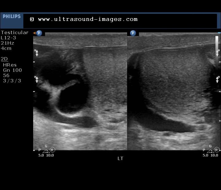


The left testes also shows tubular ectasia of the rete testes- in the 3-D ultrasound image below:


Visit: http://www.ultrasound-images.com/scrotum/#Hydrocele-%20B-mode%20grey%20scale%20and%203-D%20images

This comment has been removed by a blog administrator.
ReplyDelete