Wednesday, March 13, 2013
Thursday, March 07, 2013
Wednesday, March 06, 2013
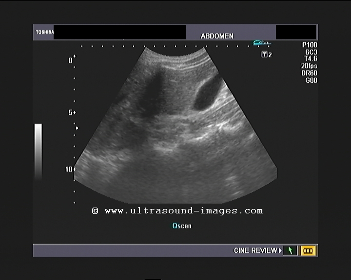
Portal cavernoma Vs portal biliopathy- a sonographic case study
This elderly male patient had generalized discomfort in the abdomen with vague pain in the epigastrium. No h/o alcoholism.
Useful reading : 1) Sonoworld article on portal cavernoma
2)Radiopedia article on cavernous transformation of the portal vein
3) MRI in portal biliopathy
4) RSNA - radiology article portal biliopathy
Useful reading : 1) Sonoworld article on portal cavernoma
2)Radiopedia article on cavernous transformation of the portal vein
3) MRI in portal biliopathy
4) RSNA - radiology article portal biliopathy
Wednesday, February 20, 2013
Severe left internal carotid artery stenosis:
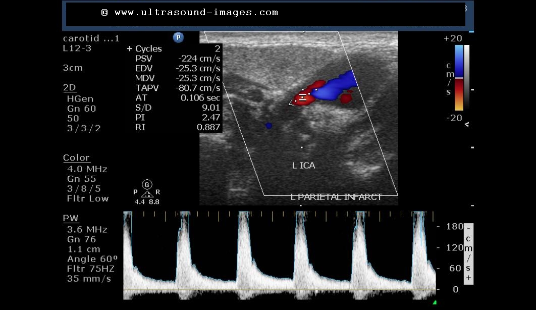
This patient has the right hemiplegia following a cerebrovascular accident involving the left parietal lobe. Color Doppler imaging of the carotid tree shows severe stenosis, 80 to 90%, involving the left internal carotid artery close to its origin from the common carotid artery.
The colour Doppler image below shows the internal diameter of the left internal carotid artery is less than 3 mm near its origin. This is definitely abnormal and shows severe stenosis.
spectral Doppler trace of the left common carotid artery appears relatively normal.
however, the left internal carotid artery shows a definite evidence of severe stenosis with peak systolic velocity reaching up to 224 cm/s suggesting greater than 90% stenosis at this level. The normal peak velocity of flow at the internal carotid artery should be less than an and 120 cm/s.
the video below shows the characteristic spectral waveform seen in this case. The characteristic high pitch sound of the Doppler waveform and high velocity flow at the site of left internal carotid artery stenosis can be appreciated.
For more on this topic: http://www.ultrasound-images.com/vascular.htm#Internal_Carotid_artery_occlusion
The colour Doppler image below shows the internal diameter of the left internal carotid artery is less than 3 mm near its origin. This is definitely abnormal and shows severe stenosis.
spectral Doppler trace of the left common carotid artery appears relatively normal.
however, the left internal carotid artery shows a definite evidence of severe stenosis with peak systolic velocity reaching up to 224 cm/s suggesting greater than 90% stenosis at this level. The normal peak velocity of flow at the internal carotid artery should be less than an and 120 cm/s.
the video below shows the characteristic spectral waveform seen in this case. The characteristic high pitch sound of the Doppler waveform and high velocity flow at the site of left internal carotid artery stenosis can be appreciated.
For more on this topic: http://www.ultrasound-images.com/vascular.htm#Internal_Carotid_artery_occlusion
Friday, February 08, 2013
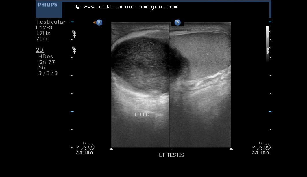
A case of painful left scrotal mass
This middle aged male patient has a left scrotal mass which is tender and painful. What do you opine?
The mass showed plenty of mobile particulate matter and is located on the upper pole of the left testis.
Answer: this is a case of left scrotal abscess with the purulent collection being within the left tunica vaginalis sac. Aetiology of this left scrotal collection appears to be tuberculous in origin. This can explain the lack of significant vascularity within the left scrotum and the left testes.
See similar lesions at: http://www.ultrasound-images.com/scrotal-infections.htm
The mass showed plenty of mobile particulate matter and is located on the upper pole of the left testis.
Answer: this is a case of left scrotal abscess with the purulent collection being within the left tunica vaginalis sac. Aetiology of this left scrotal collection appears to be tuberculous in origin. This can explain the lack of significant vascularity within the left scrotum and the left testes.
See similar lesions at: http://www.ultrasound-images.com/scrotal-infections.htm
Saturday, January 26, 2013
what is your diagnosis of this cystic lesion?
This middle aged woman had a history of fibroid of the uterus diagnosed a few years before. Now she presented with discomfort in the pelvic region. Ultrasound images of the uterus taken both transabdominally and via the transvaginal route are shown below:
Firstly, determine the location of this lesion and then try to arrive at a diagnosis based on the history and the ultrasound pictures shown above.
Answer: this cystic lesion is a cystic degeneration of an earlier or pre-existing fibroid of the uterus.
Cystic degeneration of the fibroid is a known medical entity and is a form of necrosis of the solid tumor matter within the fibroid.
For more on fibroids: http://www.ultrasound-images.com/uterus.htm
Also: http://cochinblogs.blogspot.in/2010/11/fibroids-of-uterus-leiomyoma.html
Also this article on cystic change in fibroid by IJRI
Firstly, determine the location of this lesion and then try to arrive at a diagnosis based on the history and the ultrasound pictures shown above.
Answer: this cystic lesion is a cystic degeneration of an earlier or pre-existing fibroid of the uterus.
Cystic degeneration of the fibroid is a known medical entity and is a form of necrosis of the solid tumor matter within the fibroid.
For more on fibroids: http://www.ultrasound-images.com/uterus.htm
Also: http://cochinblogs.blogspot.in/2010/11/fibroids-of-uterus-leiomyoma.html
Also this article on cystic change in fibroid by IJRI
Subscribe to:
Posts (Atom)