Wednesday, December 20, 2006

Ultrasound scan of rupture of uterus:


Rupture of the uterus during delivery is a known complication of pregnancy. The commonest cause is vaginal delivery after a caesarean section. Other causes include, previous surgery for tumors like fibroids and also prior classical caesareans, where the incision was placed in the upper uterus. Labor inducing medications can also cause rupture. I present ultrasound images of uterine rupture with severe hemorrhage on my ultrasound gallery at: http://drjoea.googlepages.com/ultrasoundgallery-uterus
On ultrasound scan, the typical findings are breach of the uterine wall, with a hypoechoic collection of blood outside the uterus. Follow up scans revealed diminishing of the size of the collection. One of the images of this case is shown here. Images courtesy of Dr. Durr-e-Sabih, Pakistan.

Monday, December 04, 2006

3-D ultrasound of animal fetuses:

Ever wondered how the fetus of an elephant, a dog or a dolphin appears on ultrasound?
Then try this link: Animal ultrasound
You dont need a diploma in radiology to identify the animal fetus :-)


Friday, December 01, 2006

The latest buzz on Teleradiology:

A recent survey by researchers in Yale University in New Haven, CT suggests that American physicians would prefer to have domestic radiology services, than having teleradiologists, based overseas, report the scans or X-rays of their patients. This survey reflects strong opposition, among a section of clinicians and radiologists, to the globalization of radiology services. While US based radiologists have a reason to feel threatened by their counterparts in Asia, because of loss of their share of the pie, clinicians are concerned about the quality of teleradiology services. More on this at the link:
Survey finds domestic teleradiology preferred by referrers
This represents insecurity and anxiety as the “dirty word” ie: “outsourcing” enters the US medical arena. But if banks, financial institutions and big businesses can do having their work outsourced to India, how long will it be before the American medics follow?
Dr. Joe Antony, MD.
Do visit:
free to view ultrasound image gallery>>http://drjoea.googlepages.com/
I also have a medical imaging blog at:http://www.healthvoices.com/taxonomy_menu/3/48

Friday, November 24, 2006

The role of Ultrasound imaging in the diagnosis of scrotal swellings:


Swellings, with or without pain, of the scrotum, are very common in males. Swellings of the scrotum are commonly due to collection of fluid within the membranes covering the testes. These are called hydrocele. Other causes of swellings include testicular tumours (growths), which may be cancerous, a hernia descending into the scrotum, congenital conditions like an additional testes, cysts etc. The clinician has a hard time differentiating these various conditions, but a couple of minutes spent doing an ultrasound scan of the scrotum is sufficient to diagnose all these. I discuss the commonest cause of scrotal swelling, namely a hydrocele or pyocele. This condition can be easily treated by the urologist or general surgeon.
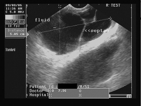
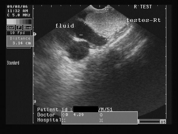
Hydrocele is an abnormal collection of fluid, usually serous, between the the two layers of the tunica vaginalis covering the testes. It may be congenital or acquired. Hydrocele is easily diagnosed by ultrasound scan. A pyocele is the same as a hydrocele, but has purulent fluid within it. Hematocele is a hydrocele with blood within the fluid and almost always is the result of trauma. Both pyoceles and hematoceles contain multiple septae (membranous partitions) within the fluid collection. At my web page: http://sites.google.com/site/drjoea/scrotum I present images of a septate collection, possibly a pyocele, and a large one at that, involving the right side of the scrotum.
DR. Joe Antony, MD.
A large free ultrasound image gallery can be found at:
http://sites.google.com/site/drjoea/home
Also visit: http://www.ultrasound-images.com/scrotum.htm
and http://www.ultrasound-images.com/acute-scrotum.htm for lots of information on sonography of scrotum.
For ultrasound study of scrotal infections visit:
http://www.ultrasound-images.com/scrotal-infections.htm

Case-2: moderately large right hydrocele/ pyocele with scrotal calculus:
This young adult male had a right testicular swelling of 3 months duration. Ultrasound images are shown below:


The ultrasound images show a large right hydrocele with particulate matter and septae. Possible infection of the hydrocele resulting in what is termed as pyocele.
Color Doppler image is shown below:


Color Doppler image shows almost normal vascularity in the right testis, ruling out orchitis.
Observe also the presence of the hyperechoic focus anteriorly, on the testicular surface- a scrotal calculus. This is literally a stone inside the scrotum, between the layers of the tunica vaginalis. Scrotal calculi can be mobile or adherent.
 

 This transverse section ultrasound video clip shows the magnitude of the problem, panning from the upper to the lower poles of the right testis.

Saturday, November 18, 2006

Sonographic diagnosis of conjoint twins:


Ultrasound Scan image (transverse section) of the conjoint twins sharing a common heart (such twins cannot survive). Images courtesy Dr. Durr-e-Sabih, Pakistan.


Post delivery view of the conjoint twins:
(below)


Conjoined twins or Siamese Twins (to the layman) are the result of incomplete division of the embryonic disc. The twins here are partially joint at either of the following points:
Head- called craniopagus
chest- thoracopagus
abdomen- omphalopagus
pelvis- ischiopagus
The incidence of such an anomaly occurring is very rare: 2 in 100000 births approximately. Fusion of the chest and abdomen is the commonest variety and is called thoraco-omphalopagus. It is associated with high mortality of the fetuses (most of the fetuses are born premature or still born). Ultrasound scan during pregnancy is very important to diagnose this condition.
Please click the link: http://drjoea.googlepages.com/obstetric-2
Here, I have presented ultrasound images of 2 different cases, with this anomaly.
Here, the twins are fused along the chest and abdomen with shared liver and heart. Such conjoined twins cannot survive. Diagnosis of conjoined twins is possible using ultrasound scan in the first trimester (first 3 months of pregnancy), but details are better visible in the 2nd trimester.
Dr. Joe Antony, MD.

free to view ultrasound image gallery>> http://drjoea.googlepages.com/

http://www.squidoo.com/conjoined_twins/
Also check my digg stories: http://digg.com/users/drjoea/dugg

My technorati link:

Technorati Profile