Interested in Ultrasound-- then visit our sister web site:
Diagnostic ultrasound gallery
You'll find loads of ultrasound images with a brief description of each case, links etc.
Thursday, September 21, 2006


The crisis facing the small clinic in India:
The small, friendly neighbourhood clinic has for long been the backbone of medical practice in India. But of late, the arrival of large hospitals or multispeciality hospitals is rapidly changing the scenario.
In Cochin, where I work, many of the smaller and even midsized hospitals are feeling the heat, as large hospitals mushroom all over the place. The situation in the field of radiology is also undergoing rapid change. Until 5 years back, independent diagnostic centers ruled the turf of radiology. There were diagnostic centers with CT scan and ultrasound facilities “under one roof”. Then MRI scan was also added to the range of services. When even that was not sufficient, pathology labs were added to these “centers”. Even large hospitals would refer cases to these diagnostic labs. But, soon, these same hospitals decided that, rather than buy the cake, they might as well make it. The result, almost all the major hospitals in town have their own complete diagnostic facilities. The situation is so bad, that some of the independent labs have shut shop or shifted their equipment to greener pastures.
So, what does the future hold for medical practice here. Obviously, many of the numerous labs (and also the smaller hospitals) in this part of the world have to adapt to the changing times; many might simply close shop. Others would merge with the large mega-hospitals, to survive. The friendly neighbourhood clinic, or lab might operate on a part- time basis, with the radiologist operating part time at a number of labs.
The trends one sees in the corporate world will be mirrored in the medical field also.
Kerala, with its high literacy rates (almost 100%) and consequent health consciousness, coupled with inflow of petro- cash (from overseas Indians in the Arab countries), has an abundance of private healthcare facilities. In fact, it is estimated, that, Kerala has one of the highest number of CT scan machines, per unit population in the world. The changes in this microcosm will reflect those in the West and elsewhere.
In Cochin, where I work, many of the smaller and even midsized hospitals are feeling the heat, as large hospitals mushroom all over the place. The situation in the field of radiology is also undergoing rapid change. Until 5 years back, independent diagnostic centers ruled the turf of radiology. There were diagnostic centers with CT scan and ultrasound facilities “under one roof”. Then MRI scan was also added to the range of services. When even that was not sufficient, pathology labs were added to these “centers”. Even large hospitals would refer cases to these diagnostic labs. But, soon, these same hospitals decided that, rather than buy the cake, they might as well make it. The result, almost all the major hospitals in town have their own complete diagnostic facilities. The situation is so bad, that some of the independent labs have shut shop or shifted their equipment to greener pastures.
So, what does the future hold for medical practice here. Obviously, many of the numerous labs (and also the smaller hospitals) in this part of the world have to adapt to the changing times; many might simply close shop. Others would merge with the large mega-hospitals, to survive. The friendly neighbourhood clinic, or lab might operate on a part- time basis, with the radiologist operating part time at a number of labs.
The trends one sees in the corporate world will be mirrored in the medical field also.
Kerala, with its high literacy rates (almost 100%) and consequent health consciousness, coupled with inflow of petro- cash (from overseas Indians in the Arab countries), has an abundance of private healthcare facilities. In fact, it is estimated, that, Kerala has one of the highest number of CT scan machines, per unit population in the world. The changes in this microcosm will reflect those in the West and elsewhere.
Sunday, September 10, 2006
The future of MRI scan- Functional MRI:
Functional MRI scans – the future of MR imaging:
The latest in imaging has arrived, in the form of Functional Magnetic Resonance Imaging or F- MRI scans.
Imaging remained largely a means to study anatomical lesions of the body, such as tumours, infections etc. But now, F- MRI is set to change all that. F- MRI is a technique that actually maps hemodynamic changes (read blood flow changes) in response to
neural stimuli or neural activity, in the brain and spinal cord. This is based on the fact that that there are changes in blood flow patterns in regions of the brain that are active; for example moving the fingers would produce more blood flow and oxygenation in the region of the brain called the motor cortex. The MR signals of the blood vary according to the degree of oxygenation. This fact is made use of to color code the brain’s activity. F- MRI can thus non-invasively detect human brain tissue activity, without the fear of radiation.
I had the chance to see a slide show, where an F- MRI scan of a patient with anxiety disorder, was displayed. Multiple areas (colored red) were seen along both cerebral hemispheres. After making the same patient relax, listening to gentle music, a repeat F MRI scan showed a near normal appearance. Thus, the role of this technique, in neurology and psychiatry is obvious. A more controversial use of the technique in comatose patients, found that in some such cases, in so called minimally conscious states,
the brain showed responses to speech and external stimuli, similar to those in normal persons. This raises many questions, indeed a Pandora’s box of legal and ethical issues regarding the conventional definition of death.
The latest in imaging has arrived, in the form of Functional Magnetic Resonance Imaging or F- MRI scans.
Imaging remained largely a means to study anatomical lesions of the body, such as tumours, infections etc. But now, F- MRI is set to change all that. F- MRI is a technique that actually maps hemodynamic changes (read blood flow changes) in response to
neural stimuli or neural activity, in the brain and spinal cord. This is based on the fact that that there are changes in blood flow patterns in regions of the brain that are active; for example moving the fingers would produce more blood flow and oxygenation in the region of the brain called the motor cortex. The MR signals of the blood vary according to the degree of oxygenation. This fact is made use of to color code the brain’s activity. F- MRI can thus non-invasively detect human brain tissue activity, without the fear of radiation.
I had the chance to see a slide show, where an F- MRI scan of a patient with anxiety disorder, was displayed. Multiple areas (colored red) were seen along both cerebral hemispheres. After making the same patient relax, listening to gentle music, a repeat F MRI scan showed a near normal appearance. Thus, the role of this technique, in neurology and psychiatry is obvious. A more controversial use of the technique in comatose patients, found that in some such cases, in so called minimally conscious states,
the brain showed responses to speech and external stimuli, similar to those in normal persons. This raises many questions, indeed a Pandora’s box of legal and ethical issues regarding the conventional definition of death.
Wednesday, August 30, 2006
HOW SAFE IS YOUR CT SCAN?
Risk of radiation during CT scans:
CT scans have become commonplace and are an essential part of investigations,
that the consultant/ doctor advises, today. However, have CT scans been taken for granted, as truly safe for the patient?
Recent studies done (http://www.fda.gov/cdrh/ct/risks.html ) in USA and Europe suggest that there is a small but significant risk of radiation induced cancer, occurring due to
CT scans. These conclusions have been made after the release of the BEIR VII report
( BEIR VII Press Release: Low Levels of Radiation May Cause Harm ).
According to the report, the National Academy of Sciences, has described a 1-in-1000 chance of developing cancer from a single radiation exposure of 10 mSv.
(mSv is a unit of measuring the amount of radiation a person receives).
10 mSv is the amount of radiation a patient receives during a single CT scan study of the
abdomen, chest or the pelvis. The study also finds that the risk in children is even
higher. 1 in every 550 children scanned would develop cancer due to the CT scan.
The recommendations of the study are that persons under 40 yrs. age should be carefully
screened to avoid unnecessary scans. This is specially true whilst scans involve the gonads.
A CT scan of the head exposes you to about 2 mSv, a scan of the chest is about 8 mSv, and a study of the abdomen and pelvis is about 10 mSv. A normal person is
exposed to 3 to 5 mSv of radiation every year (ie: radiation from soil and atmosphere).
The real problem arises when a person (especially those under 40 and children) are
subjected to repeated scans, with doses exceeding 50 mSv.
Conclusion: Be judicious in the use of CT scans. Though there are definite risks
involved in repeated scans, they are to be carefully weighed against the numerous diagnostic benefits this tool provides.
CT scans have become commonplace and are an essential part of investigations,
that the consultant/ doctor advises, today. However, have CT scans been taken for granted, as truly safe for the patient?
Recent studies done (http://www.fda.gov/cdrh/ct/risks.html ) in USA and Europe suggest that there is a small but significant risk of radiation induced cancer, occurring due to
CT scans. These conclusions have been made after the release of the BEIR VII report
( BEIR VII Press Release: Low Levels of Radiation May Cause Harm ).
According to the report, the National Academy of Sciences, has described a 1-in-1000 chance of developing cancer from a single radiation exposure of 10 mSv.
(mSv is a unit of measuring the amount of radiation a person receives).
10 mSv is the amount of radiation a patient receives during a single CT scan study of the
abdomen, chest or the pelvis. The study also finds that the risk in children is even
higher. 1 in every 550 children scanned would develop cancer due to the CT scan.
The recommendations of the study are that persons under 40 yrs. age should be carefully
screened to avoid unnecessary scans. This is specially true whilst scans involve the gonads.
A CT scan of the head exposes you to about 2 mSv, a scan of the chest is about 8 mSv, and a study of the abdomen and pelvis is about 10 mSv. A normal person is
exposed to 3 to 5 mSv of radiation every year (ie: radiation from soil and atmosphere).
The real problem arises when a person (especially those under 40 and children) are
subjected to repeated scans, with doses exceeding 50 mSv.
Conclusion: Be judicious in the use of CT scans. Though there are definite risks
involved in repeated scans, they are to be carefully weighed against the numerous diagnostic benefits this tool provides.
Monday, July 10, 2006
CLICK FOR FREE MUSIC

Looking for free music downloads, try this internationally acclaimed site:
Click Here To Download Free Music and Movies
Scan your computer hard drive for malicious software, adware, scumware etc.
you might have picked up from the internet.

Thursday, May 04, 2006
Dr.Anatoly--a radiologist and an artist

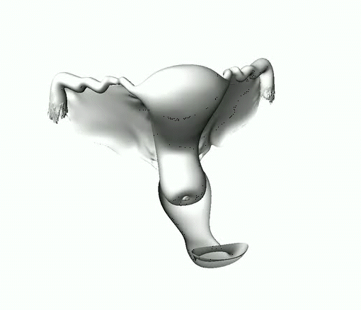
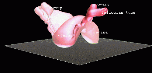
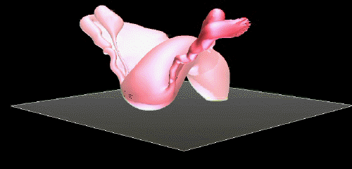
My good friend, Dr. Anatoly, from Kiev, Ukraine,
feels the human body and its insides
are a work of art to be perfected through 3D
graphics as he demonstrates so well in these pictures.
They show the female reproductive organs in
3Dimensions. Unfortunately, this site does not
allow moving 3D- animations which is what these are.
Subscribe to:
Posts (Atom)