Friday, November 26, 2010

Causes of intertility in men and women:

Among the causes of infertility in women are diseases of the uterus and ovaries:
This middle aged lady has a very small uterus (called hypoplasia of the uterus). Such a uterus is incompatible with the ability to conceive and have a child. The ultrasound video clip below shows a sagittal (midline) section through the uterus. It was obtained via the transvaginal route. The vagina itself was sufficiently large enough to insert the transvaginal probe.
 
The inner lining of the uterine cavity- the endometrium appears markedly thinned, and again this is incompatible with conceiving a child. 

 
The ultrasound video clip below is a transverse section through the uterus of the same patient:
In men, similarly, absence of or hypoplasia (very small) of parts of the reproductive system can result in infertility:
This male patient underwent sonography of the prostate and seminal vesicles via the transrectal route:
Ultrasound video clip shows normal sized prostate-


Observe the TRUS video clip as we pan the probe from the upper most part of the prostate showing the clear absence of the seminal vesicles and the vas deferens (agenesis) on both sides. This type of congenital absence of an important part of the route through which sperms pass from the testes to the penile urethra will result in total absence of spermatozoa in the ejaculated semen. This condition is called a azoospermia in the male.

Other and more common causes of infertility in men-
Perhaps one of the commonest cause of male infertility is a condition called varicocele.
Here, there is a dilatation of the veins around the testes, inside the scrotum. These veins are called the Pampiniform veins, and are responsible for draining blood from the testes. Due to decreased efficiency of the draining process, these veins swell and blood collects within these vessels resulting in increased temperature within the scrotum and the testes. The testes then lose some of their spermatogenic (sperm producing) functions. Depending on the degree of varicocele (grade of varicocele), the man may suffer from infertility.  
Color Doppler Ultrasound can help in diagnosing this condition; see this link for more on this topic:
Diseases of the testes and the epididymis (a small structure next to the testis) can cause poor or impaired production of sperms. This can result in infertility. One such common condition is an inflammatory disease called orchitis and epididymo-orchitis. (see: http://www.ultrasound-images.com/scrotum.htm#Epididymitis
Another cause of impaired spermatogenesis (sperm formation) is chronic or past infection of the testes resulting in atrophy (shrinking) of the testes. This happens in mumps inflammation of the testes, a condition called mumps orchitis. See : http://www.ultrasound-images.com/scrotum.htm#Atrophy_of_testis
This condition causes decreased function of the testes with small shrunken testes. Atrophy of the testes is usually irreversible.
Another condition that involves another part of the male reproductive system is prostatitis or inflammation of the prostate. The prostate helps to form a bulk of the fluid in the semen. Inflammation of the prostate is common and can impair the production of semen and its release during ejaculation. This color Doppler video clip shows a severely inflamed prostate:


This prostate was visualized via the transrectal route. The color Doppler mode shows massively increased blood flow in the prostate (hyperemia), a hall mark of prostatitis. This condition of the prostate can be corrected to a large extent using medical treatment.


The above still color Doppler image shows the TRUS (transrectal ultrasound) view of the same prostate.

Sunday, November 07, 2010

Fibroids of the uterus (leiomyoma):

Fibroids are the commonest tumors of the uterus and have a reported incidence of at least 25 to 30 % in the female population. Uterine fibroids are benign tumors (lumps) of the uterine musculature (called smooth muscles) and have varying prognosis from slowly growing tumors to those that may actually regress (become smaller). A small percentage of fibroids of the uterus may actually undergo malignant transformation (become cancerous); these cancerous tumors are called leiomyosarcoma. Another aspect about a fibroid is that this uterine tumor may be single or it may be multiple.
What are the symptoms of a uterine fibroid?
The earliest symptoms of a fibroid can be pain the lower abdomen (the pelvis) of the woman (usually after the age of 30). The other symptoms of uterine fibroids include dysmenorrhea (pain during menses), increased frequency or amount of bleeding during menses (polymenorrhea or menorrhagia) and other menstrual abnormalities. Other symptoms include those produced by pressure of the fibroids of the uterus on the adjacent organs in the female pelvis including the urinary bladder (increased frequency of urination and obstructed urination). Pressure on the rectum may cause constipation in women where the uterine mass (tumor/ fibroid) arises from the posterior (back) of the uterus. Fibroids are also a common cause of infertility in women.
 So how does one diagnose fibroid of the uterus?
If a woman has one or more of the symptoms mentioned above, she should consult the doctor/ gynecologist immediately. Per abdominal examination (palpation) of the pelvis can sometimes help detect a relatively larger fibroid. In addition, a per vaginal examination may also help to detect a lump of the uterus. In any case, the safest and best method to diagnose a fibroid of the uterus is ultrasound examination. The sonologist or radiologist conducts a transabdominal examination of the pelvis (on a full bladder) to visualize the uterine mass. The uterine tumor is seen from various angles to determine its size, number, location and pressure effects on the adjacent bladder or rectum.
Here is some examples of what a uterine fibroid looks like on ultrasound imaging:
There are commonly three types of fibroid based on ultrasound imaging:
a) Intramural- the fibroid is within the wall of the uterus.
b) Subserosal- the fibroid is just below the outer covering serosa of the uterus.
c) Sub mucous- the fibroid is just below the inner lining of the uterus (the endometrium).


Image below shows multiple fibroids:


This ultrasound image shows a fibroid extending from the subserous to the intramural location of the uterus-


What does the sonologist or radiologist see in this ultrasound image of the subserosal fibroid? The uterine fibroid is pressing upon the anteriorly (front) located urinary bladder. The mass or tumor is located to the right side of the uterus and extends from the intramural to the subserosal location of the upper part of the uterus (called the fundus).
See another ultrasound picture of the same case:


The uterine fibroid also appears to compress upon the inner lining of the uterus (EM= endometrium).(FIB= fibroid; BL= bladder; UT= uterus). Thus this patient is likely to have a number of complaints- from bladder compression to irritation of the inner lining (endometrium) of the uterus with resultant menstrual problems.
Broad ligament fibroid:
In rare cases the uterine fibroids may be located to either side of the uterus- the so called broad ligament fibroids- see the ultrasound pictures/ ultrasound video clips of one such case of broad ligament fibroid:
The uterine tumor in this case is seen in the left broad ligament of the uterus. Such a broad ligament fibroid may interfere with the function of the left fallopian tube (the duct that transmits the ovum from the left ovary to the uterine cavity). Hence such a patient might have problems conceiving or may be infertile.
See this ultrasound video clip of the same case (left broad ligament fibroid):

Pedunculated fibroid:
The next case we shall discuss is another relatively rare entity called the pedunculated fibroid of the uterus. Here, the fibroid in the uterus is connected to the uterus by a short or sometimes long pedicle or stalk. See the ultrasound images of one such case of pedunculated fibroid of the uterus:

In this patient, the uterine fibroid is seen extending just above the fundus (top) of the uterus and is connected to it by a short stalk. It is almost like a floating tumor suspended from the main body - the uterus by a stalk or cord. Such pedunculated uterine fibroids can cause severe pain due to torsion of the uterine tumor mass (twisting of the stalk) resulting in cutting off of the blood supply to the tumor from the uterus.
Have a look at these ultrasound video clips of the pedunculated fibroid described above:


Such an event resulting in twisting of the stalk of the pedunculated fibroid can be a medical emergency and urgent surgery might be required to remove the mass immediately.


This color Doppler ultrasound video clip shows the stalk is very vascular (ie: a number of blood vessels are seen between the pedunculated uterine fibroid and the fundus of the uterus. For more details of the various other types of fibroids of the uterus and ultrasound images of each case, visit:


Case-2 (pedunculated fibroid of uterus):
This is another example of a large fibroid with a stalk connecting the tumor (6 x 7 cms.) to the fundus of uterus via a stalk or peduncle. The peduncle measures about 2 cms. in thickness. I managed to visualize the peduncle by applying pressure onto the area between the fibroid and the fundus of the uterus. See the ultrasound video below:


Color Doppler video clip (above) shows multiple blood vessels within the short and relatively thick pedicle connecting the fundus to the fibroid.
This is how the mass (fibroid) and pedicle appeared on still images (see below):


 Observe the presence of a second fibroid within the lower part of body of the uterus in these sagittal sections of the uterus. This fibroid appears intramural in location.
It is beyond the scope of this article to describe in detail the other methods of diagnosing tumors of the uterus. However, it must be added that CT (CAT) scan imaging and MR imaging can also offer additional information on  fibroids of the uterus, in certain cases.
For information on treatment of fibroid of the uterus visit:
Degenerative changes in fibroid:
This case of a large fibroid with a most unusual appearance:
   
This is a sagittal section of the huge mass in the uterus. Note the markedly inhomogenous appearance of the mass. The ultrasound video clip below shows a transverse section of the mass:


Now, the color doppler video of the tumor (fibroid) of the uterus:


For more images and details of this case see:
http://www.ultrasound-images.com/uterus/#Types%20of%20fibroid

Multiple fibroids:
Uterine fibroids can very often be multiple. This middle aged lady has 3 fibroids seen in this sagittal section ultrasound video clip of the uterus:
Of these, the one labeled as (1)- towards the fundus, shows degenerative changes- it is hypoechoic, inhomogenous, suggesting possible cystic changes within it.
Another video clip shows the presence of all 3 fibroids in this uterus:

Sunday, October 10, 2010

Ultrasound Imaging of the head of neonatal femur and humerus

High resolution Ultrasound images of the normal head of femur in neonate:
 The above image shows the head of the left femur in a 3 day old neonate. Note the full ball of the head of the femur in this transverse section of the femoral head inside the left hip joint. 
 Another neonate- the head of femur is seen a round, hypoechoic structure due to its cartilaginous nature. Note the coarse appearance of the femoral head.


Ultrasound image of the right femoral head (hip joint) - all the above images show the normal head within the acetabulum (socket of the hip joint).                                                                                                                                 
This (above) ultrasound image shows the normal head of the left humerus (in neonate). As in the case of the femoral head images, the head of the humerus is cartilaginous at this young age- hence the hypoechoic, grainy or coarse echotexture. But the humerus shows one major difference- the head forms a "half ball" -a shallow ball and socket joint (the shoulder joint). In infective arthritis of these joints, there would be widening of the joint space with collection of synovial fluid within the joint cavity. Ultrasound is an excellent method of imaging the neonatal hip and shoulder joints.


 The above 3 high resolution (7- 8 Mhz transducer frequency) show the normal right neonatal shoulder joint, from different angles. The transverse section image shows the shallow nature of the glenoid cavity. Sonography of the shoulder joint in the neonate is useful in detecting tendency for dislocation in a baby and ruling out conditions like shoulder dysplasia in infants. By detailed ultrasound imaging one can detect glenoid dysplasia in infant shoulder (a clear indicator of potential shoulder dislocation). This condition is coomon in neonates/ infants with obstetric brachial plexus injury.  

Friday, August 20, 2010

Color Doppler ultrasound videos of 3rd trimester pregnancy:

Color Doppler ultrasound is an essential part of the imaging or ultrasound scan of the fetus, even in the 3rd trimester. the basic Doppler imaging of the fetus includes study of the fetal MCA or middle cerebral artery, the fetal ductus venosus and the umbilical artery. Of these, Doppler imaging of the ductus venosus is the most difficult. Here we have some normal Triplex Doppler videos of these 3 main vessels of interest to the sonographer or radiologist. All the Color Doppler videos below show video and accompanying audio.
Color Doppler and spectral waveform of the fetal middle cerebral artery:
Here is the second video of the MCA:
                                                                             This Color Doppler video clip demonstrates the exact location of the middle cerebral artery (arrow) among the major vessels of the fetal brain.  
Yet another demonstration of the fetal middle cerebral artery (arrow pointing at the vessel) :
 
Due to fetal head movement and respiratory movements the arrow points slightly lower than the actual position of the fetal MCA.

                      Note the high velocity, high resistance flow in this normal fetal middle cerebral artery, with almost no diastolic flow that is very striking about this artery.
 The other important fetal vessel that is studied here is the umbilical artery. This spectral Color Doppler waveform video clip shows the normal umbilical artery. The normal umbilical artery shows low resistance flow with high diastolic velocity, which is the opposite of what is seen in the fetal MCA.                                                                                                           
                                                                                 The next vessel to be studied in this normal fetus is the most elusive as we mentioned earlier and is the fetal ductus venosus. This color Doppler video /spectral waveform shows the normal ductus venosus with low resistance flow with typically 3 waves seen in spectral display- the S, D and A waves. This vessel is short and disappears from view unless the clinician has a steady hand on the probe and both mother and fetus cooperate.                                                                                                                                                     

                                                                              Here is another attempt at the normal ductus venosus in this 3rd trimester fetus:
                                                                            The Nemio-XG ultrasound machine which I have used for this study has an "update" button which helps to fine tune the spectral video display. This update button has been activated to the later half of this ductus           venosus video. The result is obvious, as the waves become smaller and more coherent.                                                                                                       Note the almost M-shape of these normal ductus venosus waves. For more on this topic visit:                                        http://www.ultrasound-images.com/fetus-general.htm for more images and case studies of abnormal fetal doppler.                 
Here is yet another spectral Doppler video tracing of a 34 week old fetus (another case):     
Often, this short vessel slips out of view due to fetal movements or maternal breathing. Hence a lot of effort is needed to get optimal color Doppler spectral tracing of fetal ductus venosus.


Saturday, August 14, 2010

Gall bladder sludge- ultrasound video clip:

This B-mode ultrasound video clip shows what is called as limy bile in the gallbladder. This organ, though distended, shows echogenic particles and also calculi (stones) of 4 to 6 mm. in its lumen. See this page for more: 


The bile in the gallbladder must be a rather thick fluid, almost jelly or putty like in consistency, given that the ultrasound video clip shows almost no motion of the debris in the gallbladder. The overall appearance and echogenicity of the gall bladder is similar to that of the liver, an appearance called hepatization.

Friday, August 13, 2010

Color Doppler ultrasound video of a ureteric jet:

 
This short Color Doppler video clip shows a jet of urine (red stream) emerging from the left ureteral orifice and sprayed into the distended urinary bladder. The jet of urine was normal in this patient. A normal urine jet from the ureter is an indication that there is no obstruction in the ureter of that side, at least not at the level of the ureteric orifice.

Wednesday, August 11, 2010

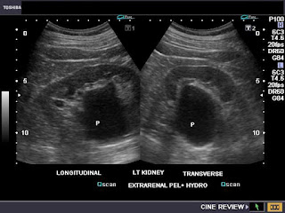
Extrarenal pelvis:


This adult patient shows a ballooning of the pelvis (P) of the left kidney (see ultrasound images above). A major portion of the renal pelvis lies outside the renal sinus, a condition called extrarenal pelvis. The transverse section image (lower image), brings out the lesion clearly. The extrarenal pelvis is a normal variant and needs no specific treatment. However, in this case, the calyces of the left kidney show mild ballooning suggesting obstructive changes which require further radiological study.
Reference: http://emedicine.medscape.com/article/1016549-overview

Sunday, August 01, 2010

Solitary cyst of fetal kidney:


This 2nd trimester fetus shows a solitary cyst of the lower pole of left fetal kidney. The cyst is small (6 mm.) with no internal debris or septae. The fetal renal cyst shows no communication withe the pelvicalyces which appear norma, thus ruling out fetal hydronephrosis. The fetal urinary bladder (BL) appears normal. The above ultrasound images are typical of uncomplicated cyst of the fetal left kidney. The important differential diagnoses in this case include renal abscess or renal hematoma, both of which seem improbable here. The other possibility is that this cyst is precursor to onset of autosomal dominant polycystic kidney disease (adult type). Polycystic kidney disease may develop in this baby during adult life. Both images are courtesy of        Dr. Ravi Kadasne, MD, UAE.
For more on this topic visit: http://www.ultrasound-images.com/fetal-urogenital.htm

Saturday, July 31, 2010

Ultrasound imaging of nuchal thickness ( nuchal translucency thickness): 11 to 13 weeks fetal scan


Ultrasound imaging of the fetus in the period from 11 weeks to 13 weeks 6 days gestational age is important to study fetal nuchal translucency or nuchal thickness. Nuchal translucency is the hypoechoic area in the subcutaneous region in the posterior aspect of the fetal neck. This hypoechoic area represents fluid collection in the nuchal region. The normal amount of fetal nuchal thickness can be upto 2 mm. (at 11 weeks). The normal nuchal translucency thickness can increase to about 2.8 mm. by 13 weeks 6 days. The above ultrasound image shows a sagittal section of the fetal neck with nuchal tranlucency or nuchal thickness of     1.8 mm. in a 11 week old fetus.  This was a normal study. Note the separate band posterior to the neck- the amnion, a membrane present around the fetus. The amnion must be separately identified from the nuchal skin to prevent wrong measurement of the nuchal thickness.


A zoomed view of the nuchal thickness in the same fetus. Note the close proximity of the amniotic membrane.


The normal nasal bone is also visualized in this ultrasound study (see image above). All above images are courtesy of Dr. Prasenjeet Singh, India.
Reference: http://www.babycentre.co.uk/pregnancy/antenatalhealth/scans/nuchalscan/
This site gives more information on this topic:
http://www.fetalmedicine.com/fmf/FMF-English.pdf
http://www.fetalmedicine.com/fmf/training-certification/certificates-of-competence/11-13-week-scan/nuchal/

Thursday, July 29, 2010

The glomus body and its tumors:

The glomus body is a unique receptor or rather a thermal sensor found mainly in the dermis of the skin. This is a strange kind of tissue, microscopic in size but with a very important function, somewhat akin to the sensors found inside the room air conditioner. It acts as a sensor detecting changes in temperature of the environment and passes this information up the nervous system resulting in shunting of blood away from the skin by vasoconstriction (shrinking the diameter of the capillaries in the skin) when the surroundings turn cool. This simple mechanism prevents loss of heat during the winter and cool weather. The opposite happens during warm weather. Perhaps we don't realize the importance of these tiny nodes in the skin until it malfunctions. Perhaps the most common pathology of this node is in the distal part of the fingers- the subungual region (the part between the finger nail and the underlying phalanx or bone). This condition is the glomus tumor, a very vascular growth, some call it a hamartoma of the glomus body.
See- http://www.ultrasound-images.com/musculoskeletal.htm , where I discuss this pathology in detail with color Doppler and ultrasound imaging of this rather poorly understood and still relatively unexplored pathology. 


 The grey scale B-mode ultrasound image above shows a typical glomus tumor, seen as a hypoechoic, soft tissue mass beneath the fingernail.
The color Doppler image above shows the vascular nature of the subungual glomus tumor in the 2nd finger.Note the erosion of the underlying bone. Both above images are courtesy of Shlomo Gobi, Israel.

Wednesday, July 21, 2010

Ureterocele with ureteric jet:


 Case-1:
Does a ureterocele prevent urine entering into the urinary bladder? This Color Doppler ultrasound image proves that ureteroceles may not always produce ureteral obstruction. Despite the large size of the outpouching of the distal end of the ureter seen in this image, there is a powerful jet of urine being pumped into the urinary bladder (the urine jet is seen as a colored column in the bladder). For more images on this topic see: http://www.ultrasound-images.com/urinary-bladder.htm
This color Doppler ultrasound image is courtesy of Dr. Prasenjeet Singh, MD, India.
Case-2:
And here is another case- a middle aged female patient with pain in the left iliac region:
Grey scale ultrasound image shows a small right ureterocele:
In addition there is a mild hydronephrosis of the right kidney.


This ultrasound video clip confirms the right ureterocele. Very often such an appearance can be mistaken for mucosal edema around a recently passed vesico-ureteric junction calculus. But this is ruled out ---


 There is no mistaking the small bulging vesicle (bubble) of the right ureterocele that measures about 9 mm. at maximum width; gradually collapsing as it empties.

This color Doppler video clip (below) shows normal flow (ureteral or ureteric jet) from the left ureteric orifice. However there is very poor flow from the right ureteric orifice (due to the ureterocele). This is in direct contrast to the case -1 above, where the ureterocele does not cause significant obstruction, despite its large size. 
 
Moral: size does not always matter when it comes to ureteroceles!!
CASE- 3:
Here is another example of a moderately large right ureterocele, with an almost normal right ureteric urine jet:
I posted this gray scale ultrasound video clip on you tube for better clarity and resolution of the video clip:


And here is the color Doppler video clip of the same case, posted by me on you tube:

This ultrasound image shows both normal kidneys, a clear sign that there is little by way of obstruction to the flow of urine on the side of the ureterocele.

Tuesday, July 20, 2010

Color Doppler video clips of umbilical vein varix

Here are some nice ultrasound and Color Doppler video clips of a 33 week fetus with intra-abdominal umbilical vein varix.


Another Doppler ultrasound video clip:


BL= fetal urinary bladder. VAR= intra-abdominal umbilical vein varix.
Note the close relation of the varix to the urinary bladder. On grey scale ultrasound, the varix may be mistaken for the fetal urinary bladder or a diverticulum from the bladder. (See ultrasound images below):


Spectral Doppler ultrasound trace shows typical venous flow in the varix, albeit turbulent. (See ultrasound picture below).
The turbulent flow within the varix is also seen in the Doppler video clips above.

Saturday, July 10, 2010

Axillofemoral graft


This elderly female patient underwent an axillofemoral graft (also called axillofemoral bypass graft) for loss of blood flow in the right femoral artery. The procedure involved connecting the right femoral artery and right axillary artery using an artificial graft tube, which was placed along the subcutaneous plane of the chest and abdomen. This patient underwent Color Doppler imaging to assess the condition and flow in the graft tube. The color Doppler image above shows the artificial axillofemoral graft tube with normal flow through it. Note the large diameter of the graft tube and location in the subcutaneous plane of the chest wall.
Reference: http://www.ajronline.org/cgi/reprint/144/5/1005.pdf
See: http://www.ultrasound-images.com/vascular.htm

Friday, July 09, 2010

Bilateral urinary calculi- an unusual case


This was a rather unusual case. This elderly male patient complained of pain in the flanks. He was suspected to have lower urinary tract obstruction (meaning a prostate enlargement). Ultrasound imaging of the kidneys (above) show bilateral moderate hydronephrosis, with more severe changes in the left kidney (right half of image). The image shows a large calculus of 2.3 cms. lodged in the left pelvi-ureteric junction (uretero-pelvic junction). But what is the cause of the right hydronephrosis?


The cause of the right hydronephrosis is now obvious- 2 calculi are seen at the right vesico-ureteric junction.


TRUS (transrectal ultrasound) examination confirms the findings. Arrows show 2 calculi in the distal most part of the right ureter (in the vesico-ureteric junction).


Despite the urinary bladder being nearly empty, the right vesico-ureteric junction calculi are seen on TRUS image (only one of the 2 calculi see in this image). The prostate itself only showed mild hypertrophy. This is the first case I stumbled upon with a VUJ (vesico-ureteric junction) and UPJ (uretero-pelvic junction) calculi in the same patient.
See this page for more: http://www.ultrasound-images.com/ureteric-calculi.htm

Saturday, June 26, 2010

Urinary bladder polyp:


Transabdominal ultrasound image showing a small, but clearly visualized solid polypoid tissue projecting into the urinary bladder close to the bladder neck. The query here is: is this mass a bladder mucosal polyp or a beak of prostatic tissue (the patient was a middle aged male with lower urinary tract obstructive symptoms).


Transrectal ultrasound image shows the polypoid mass of 12 x 5 mm. size, emanating from the bladder mucosa close to the prostate. Color Doppler ultrasound shows considerable flow within the urinary bladder polyp. This amount of vascularity is highly suggestive that this mass of tissue is a vascular mucosal polyp and not an enlarged median lobe of prostate.
For more on this see: http://www.ultrasound-images.com/urinary-bladder.htm
Also see: http://www.mdguidelines.com/bladder-polyps-benign

Wednesday, June 23, 2010

Large sebaceous cyst in unusual location


This sebaceous cyst was huge, at almost 7 x 5 cms. and located on the buttock of this elderly male patient. The ultrasound image shows a typical hypoechoic, solid, mildly inhomogenous appearance. The patient presented with complaints related to the prostate, ignoring this large epidermoid cyst.


Color Doppler image of the same cyst shows no vascularity within the mass. A sebaceous cyst contains oily, gel like semisolid material that has been unable to escape. If infected, this can turn into an abscess.
See:
http://www.ultrasound-images.com/musculoskeletal.htm
for more ultrasound images and description of sebaceous cysts.

Tuesday, June 22, 2010

Sludge balls in the Gall bladder:


Found these two echogenic masses in the gall bladder in a young, adult female patient. The question was: are these GB polyps or sludge balls. Change in posture caused them to move.
So, that ruled out polyps. Ultrasound image above shows the sludge balls in the gall bladder (arrows).


Power Doppler image (above) shows no flow within the gall bladder lesions- that again supported my inference that these were not gall bladder polyps.


High resolution ultrasound image (high frequency transducer) gave a good close up view of the gall bladder with the two sludge balls almost "kissing" each other... ;-)
See: http://www.ultrasound-images.com/gall-bladder.htm
for more on this topic.