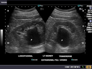
This adult patient shows a ballooning of the pelvis (P) of the left kidney (see ultrasound images above). A major portion of the renal pelvis lies outside the renal sinus, a condition called extrarenal pelvis. The transverse section image (lower image), brings out the lesion clearly. The extrarenal pelvis is a normal variant and needs no specific treatment. However, in this case, the calyces of the left kidney show mild ballooning suggesting obstructive changes which require further radiological study.
Reference: http://emedicine.medscape.com/article/1016549-overview


I just turned one of these in and called it hydronephrosis, happy to place my calipers on both sides to show how big it was. I got the Rad report back and found out how wrong I was. That's what led me here (google search).
ReplyDeleteThanks for posting.
Glad you found my blog interesting Orange Jeep.
ReplyDeleteThanks for your posting, this helps with my study tremendously !!! Thank you
ReplyDelete