I have published a nice e-book on scrotal calculi.
It is actually an ultrasound atlas of high resolution images of calcifications/ scrotal pearls.
Check this link: https://www.regnow.com/softsell/nph-softsell.cgi?item=16637-4
This e-book is in MS Power Point format...
In case you wish to get the e-book in PDF format gp to this link:
https://www.regnow.com/softsell/nph-softsell.cgi?item=16637-6
All you need to open this e-book on scrotal calculi in PDF format is adobe reader (a free download from www.adobe.com
Friday, December 18, 2009
Sunday, December 06, 2009
Sonography of penile AVM:

This ultrasound/ Color Doppler image shows multiple vessels in the glans penis and adjacent part of the penis in a middle aged male patient. This patient presented with post coital bleeding from the penis. Such a case and Color Doppler study presenting such appearances are either hardly available or known in medical literature. I have presented more Ultrasound and Color Doppler images of this very rare case at: http://www.ultrasound-images.com/penis.htm
The images show the presence of both arterial and venous flow pattern in the mesh of of vessels
in the glans penis. This is a typical appearance of arterio-venous malformation of the penis.
Tuesday, November 24, 2009
Some neat ultrasound videos of normal fetal heart
These short sonographic videos of the fetal heart show the 4 chambers clearly.
(LA= left atrium, RA= right atrium, LV= left ventricle,
RV= right ventricle).
These ultrasound video clips show the beating heart of a late 3rd trimester fetus (34 weeks gestational age).
For images, video clips of fetal heart see this link:
http://www.ultrasound-images.com/normal-anatomy.htm
For ultrasound images of fetal heart pathology and congenital anomalies of the fetal heart click this link:
http://www.ultrasound-images.com/fetal-heart.htm
Saturday, November 14, 2009
New additions to my website:
Just added some nice ultrasound images and links on renal cell carcinoma to my web site at:
http://www.ultrasound-images.com/kidneys.htm
Also an interesting addition to a rare congenital anomaly in a 2nd trimester fetus:
see- http://www.ultrasound-images.com/fetal-face-and-neck.htm
The fetal face shows a proboscis with hypotelorism, part of a series of anomalies in this pregnancy.
Joe.
http://www.ultrasound-images.com/kidneys.htm
Also an interesting addition to a rare congenital anomaly in a 2nd trimester fetus:
see- http://www.ultrasound-images.com/fetal-face-and-neck.htm
The fetal face shows a proboscis with hypotelorism, part of a series of anomalies in this pregnancy.
Joe.
Friday, November 13, 2009
Fibroadenoma and fibroadenosis of breast:

On http://www.ultrasound-images.com/breast.htm
I have just posted some nice sonographic images of a rather gray area in the field of breast imaging. There is almost no radiological literature on the subject of fibroadenosis of the breast, both on the internet and in print. At the link above are ultrasound images of both fibroadenoma and fibroadenosis of breast. What I have gathered is that fibroadenosis is a more or less "normal" nodularity of the breast which is usually self limiting (can disappear over a period of a few weeks to months). Clinically fibroadenosis may cause a little tenderness in the breast, with or without palpable nodules (which may be visualized on sonography). However, fibroadenoma of breast is a well established clinical and radiological entity. Fibroadenoma presents as a palpable (often "slippery") small nodule, well seen on ultrasound.
Here is a typical image of fibroadenoma.... (see above).
Thursday, September 17, 2009
Another case of ectopia cordis:
This is one of the best cases of ectopia cordis I have seen. The short ultrasound (sonographic) video of ectopia cordis shows the pulsating heart, outside the thoracic cavity, in both B-mode and Color Doppler imaging. Ultrasound video courtesy of Dr. Dilraj Gandhi, MD, India.
Thursday, August 27, 2009
Placental calcification
Calcification of placenta is a sign of normal degenerative changes taking place in the placenta. This usually happens in the 3rd trimester and is usually of little or no clinical significance. However, if seen earlier (2nd trimster) it might signify placental aging and abnormal degenerative changes. In the ultrasound video attached, there is extensive calcium deposition of the placenta on the maternal surface, (or basal part). Such calcific changes of the placenta do not indicate any placental pathology. See: sonography of placenta for more images and cases.
Saturday, August 22, 2009
Hydrocephalus in fetus: aqueductal stenosis:

Hydrocephalus is a common disease during intrauterine life. Almost 20 % of fetal hydrocephalus is caused by aqueductal stenosis. In the ultrasound image and sonographic video of the fetal head, above, the lateral ventricles and the upper part of the aqueduct of Sylvius (or cerebral aqueduct) are markedly dilated. The 4th ventricle does not appear to be enlarged suggesting obstruction to CSF flow at the level of the cerebral aqueduct. This is called aqueductal stenosis. Other causes of hydrocephalus include Dandy Walker malformation. Ultrasound image and video of aqueductal stenosis are courtesy of Dr. Martin Horenstein, Argentina.
For more such images of fetal brain see: http://www.ultrasound-images.com/fetal-brain.htm
Wednesday, July 01, 2009
Sonography in cervical insufficiency:

This ultrasound image of gravid uterus shows the cervix measuring 1.1 cms. in length (shortening) with dilatation (3mm.) of the internal os. The image was taken using the transvaginal route, using a Toshiba Xario ultrasound system. Dilatation with effacement of the cervix are diagnostic of cervical insufficiency. The management in this case would mean cervical cerclage.
See: Clinical aspects of cervical insufficiency (free article)
Image courtesy of Dr. Gunjan Puri, India.
From the virtual 3D image to real life size models of the fetus
Just saw this interesting link: http://www.dailymail.co.uk/sciencetech/article-1195703/The-stunning-new-technology-allows-parents-hold-life-size-model-unborn-child.html
It seems a PhD student Jorge Lopes has found a method to create 3D life size plaster models of the fetus as seen on 3D Ultrasound and MRI imaging. The replicas can help the mother identify and bond more intensely with the baby in the womb. Maybe, in the not too distant future, we may see Obstetricians and radiologists prepare plaster models, as easily as thermal print images of the fetus!!
It seems a PhD student Jorge Lopes has found a method to create 3D life size plaster models of the fetus as seen on 3D Ultrasound and MRI imaging. The replicas can help the mother identify and bond more intensely with the baby in the womb. Maybe, in the not too distant future, we may see Obstetricians and radiologists prepare plaster models, as easily as thermal print images of the fetus!!
Saturday, June 13, 2009
An aften ignored setting during sonography:


One of the most prominent settings control on any ultrasound machine is the gain knob. Yet, this happens to be one of the most unused. This knob in conjunction with the TGC or Depth gain is very useful. A good example is the female child. Here, transabdominal ultrasound to image the ovaries and small uterus can be a real pain. But lower the gain or Depth gain, and you should be able to see the ovaries, even if very small. It works in most cases.
Observe the images of the thyroid here:
With high gain settings, the margins of the thyroid and fine detail are lost. With too low gain, most of the thyroid becomes poorly visible. Optimal gain settings show both internal and marginal details. Another observation: when Color Doppler imaging is switched on, the overall gain goes down.. the gain has to be increased to compensate for this.
Tuesday, June 09, 2009
Sonography of fetal kidneys with Autosomal recessive Polycystic kidney disease

This fetal kidney shows typical features of ARPKD (autosomal recessive polycystic kidney disease) also called infantile polycystic kidney disease. This congenital disease of the kidney can be present from as early as fetal stage and may be detected as late as in childhood.
The fetal kidneys in this case show minute cysts with grossly hyperechoic kidneys.
Read more at: http://www.ultrasound-images.com/fetal-urogenital.htm
Saturday, June 06, 2009
Sedimenting echoes in urinary bladder:

Sediment producing echogenic debris in the urinary bladder can be caused by a number of factors.
Excessive amounts of Phosphate crystals in the urine is one cause. It can also be due to pyuria or pyogenic material in the urine secondary to urinary tract infection; sometimes it may be caused by hematuria (blood in urine) or chyluria following filariasis. Other causes include uricosuria (uric acid excess) or increase in oxalate crystals. This ultrasound image shows debris in the distended urinary bladder, gravitating to the dependent part.
This ultrasound image is courtesy of Dr. Ravi Kadasne, UAE.
For more sonographic images of the urinary bladder visit: http://www.ultrasound-images.com/urinary-bladder.htm
Case-2:
Here is a nice real time B-mode ultrasound video clip showing freely mobile particles in the urinary bladder:
This patient has active urinary tract infection with cystitis. The particulate matter in the urinary bladder are the result of debris- both pyogenic material and phosphate crystals floating within the urine.
This is a still image of the same case (as the video above):
Fine particles are seen throughout the urine in the bladder.
Thursday, April 30, 2009
Ultrasound video of ectopia cordis
This sonographic video shows a fetus with severe kyphoscoliosis and pulsating fetal heart lying almost outside the fetal thorax. Note the close relation between the fetal heart and fetal bladder.
The fetal abdomen shows almost no contents. The aborted fetus showed omphalocele with ectopia cordis. There was only a thin transparent membrane overlying the fetal heart, lying almost entirely outside the thorax.
The fetal abdomen shows almost no contents. The aborted fetus showed omphalocele with ectopia cordis. There was only a thin transparent membrane overlying the fetal heart, lying almost entirely outside the thorax.
Saturday, April 18, 2009
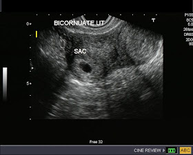
Bicornuate uterus with pregnancy


These transvaginal sonographic images show a bicornuate uterus with an early gestation sac in the right horn (cornu). Ultrasound images also show decidual tissue in the left horn. These patients can have normal pregnancy to full term, but must be carefully followed, as some may undergo a miscarriage.
Images courtesy of Dr. Jaydeep Gandhi, Mumbai, India.
Reference: http://www.ultrasound-images.com/early-pregnancy.htm
(more images and details)
