
Swellings, with or without pain, of the scrotum, are very common in males. Swellings of the scrotum are commonly due to collection of fluid within the membranes covering the testes. These are called hydrocele. Other causes of swellings include testicular tumours (growths), which may be cancerous, a hernia descending into the scrotum, congenital conditions like an additional testes, cysts etc. The clinician has a hard time differentiating these various conditions, but a couple of minutes spent doing an ultrasound scan of the scrotum is sufficient to diagnose all these. I discuss the commonest cause of scrotal swelling, namely a hydrocele or pyocele. This condition can be easily treated by the urologist or general surgeon.
Hydrocele is an abnormal collection of fluid, usually serous, between the the two layers of the tunica vaginalis covering the testes. It may be congenital or acquired. Hydrocele is easily diagnosed by ultrasound scan. A pyocele is the same as a hydrocele, but has purulent fluid within it. Hematocele is a hydrocele with blood within the fluid and almost always is the result of trauma. Both pyoceles and hematoceles contain multiple septae (membranous partitions) within the fluid collection. At my web page: http://sites.google.com/site/drjoea/scrotum I present images of a septate collection, possibly a pyocele, and a large one at that, involving the right side of the scrotum.
DR. Joe Antony, MD.
A large free ultrasound image gallery can be found at:
http://sites.google.com/site/drjoea/home
Also visit: http://www.ultrasound-images.com/scrotum.htm
and http://www.ultrasound-images.com/acute-scrotum.htm for lots of information on sonography of scrotum.
For ultrasound study of scrotal infections visit:
http://www.ultrasound-images.com/scrotal-infections.htm
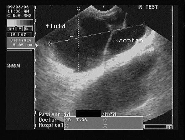
Case-2: moderately large right hydrocele/ pyocele with scrotal calculus:
This young adult male had a right testicular swelling of 3 months duration. Ultrasound images are shown below:


The ultrasound images show a large right hydrocele with particulate matter and septae. Possible infection of the hydrocele resulting in what is termed as pyocele.
Color Doppler image is shown below:

Color Doppler image shows almost normal vascularity in the right testis, ruling out orchitis.
Observe also the presence of the hyperechoic focus anteriorly, on the testicular surface- a scrotal calculus. This is literally a stone inside the scrotum, between the layers of the tunica vaginalis. Scrotal calculi can be mobile or adherent.

No comments:
Post a Comment